


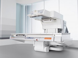
What is Fluoroscopy?
Radiologists use Fluoroscopy to obtain “live” X-Ray images of many body systems, including bones and joints, as well as solid organs such as the kidneys, urinary tract, bladder, lower digestive tract, and small and large intestines. Often the radiologist will use a contrast agent to outline other structures. Because the x-ray images are “live”, the technologist is able to capture motion, creating an x-ray “movie”.
What Are Some Common Fluoroscopic Procedures?
- Barium Enema
- Upper GI Series
- Image-guided anesthetic or contrast injections into joints or the spine
- Hysterosalpingography
- Video Swallows
What to Expect?
A PENRAD Imaging scheduler will give you instructions specific to your exam at the point of scheduling. Some fluoroscopy studies require a contrast agent, which the technologist will explain in detail. During your exam, for the best quality images, it is ideal for you to be as still as possible. The technologist will be with you for the duration of the procedure to make you comfortable and answer any questions you may have.
After the Exam
Once the test is completed, you are free to carry on with your normal activities. A board-certified radiologist will interpret your results and provide a formal report to your healthcare provider.
Recent Comments