


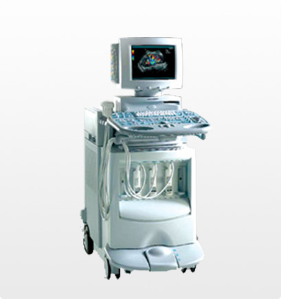
Ultrasound (also known as sonography) is a diagnostic tool using high-frequency sound waves to create images. Ultrasound is an excellent diagnostic tool to evaluate tissues, organs, and vasculature. Ultrasound cannot evaluate bone or structures that contain air. Therefore, lungs, GI tract, small bowel, and colon are not evaluated.
What to Expect
Unless instructed otherwise, many ultrasounds do not require special preparation. However, if the ultrasound is ordered to examine your abdomen and pelvic region, you may be asked to fast prior to the exam, and/or arrive at your appointment with a full bladder.
During the Exam
Our ARDMS-certified sonographer will ask you to lie on the examination table next to the ultrasound equipment. The sonographer will apply a clear water-soluble gel to the part of the body to be examined. After applying the gel, the sonographer will guide the transducer – a hand-held instrument – across your skin. The images will appear on the ultrasound monitor. Exams are typically 15-60 minutes, depending on the exam type.
After the Exam
Upon completion, the sonographer may ask you to remain in the department while reviewing the ultrasound pictures. The radiologist may also order additional images. Once the test is complete, you are free to leave and carry on with your normal activities. A board-certified radiologist will interpret your results and provide a formal report to your healthcare provider.
Recent Comments